DOWNLOAD OPHTHALMOLOGY TOOLBAR

الجمعة، رجب ٠٨، ١٤٢٩

عدسات إلكترونية ستسبق الصورة إلى عينيك قبل أن تراها

عدسات إلكترونية ستسبق الصورة إلى عينيك قبل أن تراها 8/7/2008
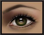
إرتدى بارباك بارفيز عدسات الإتصال في معمله والتي أحتوت على دارة إلكترونية والتي ظهرت وكأنها قطعة من عالم الخيال العلمي ,وسيقوم الأن بإضافة صمامات ضوئية متناهية الصغر تسمى (LEDs),والتي ستساعد على تحويل الطراز الأول من عدسات الإتصال إلى عدسات أكثر تعقيد تعرض أدق الأشياء التي يحتمل رؤيتها .
يعكف البرفسور بارفيز حاليًا على ما يسمى بالتجميع الذاتي أو تقنية النانو الحيوية أو إبتكار النانو والنظام الميكانيكي الإلكتروني المتناهي الصغر، فقد قام بإختراع جهاز إلكتروني متناهي الصغر واستخدم فيه تقنية النانو وتقنية إعادة التجميع المتناهية الصغر، ومن ثم قام بعملية تكامل لها إلى قطعة بوليمر أو زجاج في عملية تسمى بالتجميع الذاتي.
كيف فكر البرفسور بإختراع عدسة إتصال تحمل انظمة الكترونية دقيقة تحاكي وظائف الكائنات الحية ؟ تخيل شخصًا بمثل هذه الأمكانيات والأبحاث والخلفية الخارقة "وأضاف"تخيل ايضًا الشخص نفسه وقد استيقظ من النوم وارتدى هذا النوع من العدسات الإلكترونية.
التطلع للمستقبل
قال البرفسور بارباك بارفيز إن إختراع مثل هذا النوع من العدسات ليس بالمهمة الشاقة فهو يفكر في طريقة عرض غير تقليدية, لعدسات إتصال مصنوعة من البوليمرات الشفافة المرنة.
وكنوع من التحدي فإن بارباك يتطلع لتركيب الشرائح الإلكترونية على قطعة البوليمر هذه والحكمة هنا كما قال بارباك ليست جعل الشيء صغيرًا وإنما لجعلة مرغوبًا وسهل الإستخدام فوجود شاشة عرض في عدسة الإتصال سيسهل عملية الإستخدام. وعزا السبب إلى أن السبب كون أحجام أجهزة الحاسب المحمول والهاتف المحمول ليست صغيرة يعود إلى شاشة العرض فلو نقلنا شاشات العرض إلى عدسة الإتصال فبإمكاننا في هذه الحالة الإستغناء عن حجم الأجهزة الكبير.
وإلى الآن اظهر البرفسور اداء عالي لشرائح الإلكترونية والصمامات المتناهية الصغر ويعود الفضل لشفافيتها وحجمها الصغير ومرونتها.
وتتكون الشرائح الإلكترونية من طبقات معدينة أقل كثافة مرتبطة بالصمامات وتعتمد تقنية إعادة التجميع المتناهية الصغر والمعروفة بالتجميع الذاتي على تأثير الأنانبيب الشعرية الدقيقة في عملية ربطها ببعضها البعض لتعطي في النهاية الشكل المبدئي لشرائح الإلكترونية .
وأكد البرفسور بارفيز أن ما تحتاج إليه الشرائح اليوم الطاقة لدعمها والخطوة الرئيسة هنا التطلع لطرق مختلفة لتمرير الطاقة والطريقة المقترحة الأولى هي نقل ذبذبات الطاقة الإشعاعية ونحتاج هنا إلى هوائي لنقل البيانات إلى عدسة الإتصال والطريقة الثانية والتي نتطلع إليها بجد دمج الخلايا الشمسية ".
وأضاف" لا تعد الطاقة مشكلة صعبة التنفيذ فعدسة الإتصال تثبت مباشرة على سطح العين قريبة جدًا من عدسة العين لتركيز عليها ولإبتكار صورة مركزة يجب علينا معالجة اشعة الضوء ويمكننا ذلك إذا استخدمنا الليزر عوضًا عن الصمامات الضوئية المتناهية في الصغر إلا أن التقنية الأخيرة الأكثر سلامة في الإستخدام ".
يفكر البرفسور في إحتمال الدمج صفوف من العدسات الفردية متناهية الصغر إلى عدسة الإتصال وافاد بأن لو كان حجم الصورة قريب جدا لحجم العدسة المتناهية الصغر فإنها ستعطي صورة حيوية بحجم 30 سم بعيدة عن السطح. والتساؤل الأهم هنا ما إذا كانت هذه التقنية مؤذية للعين ؟ لأنها بذلك ستفتح افق اوسع للإتصال الوظيفي اللاسلكي في عدسات الإتصال هذه والتي سيكون لها استخدامات متعددة مستقبلاً.
ومع فكرة إنطلاق هذا الإختراع تلقى البرفسور بارفيز الكثير من الرسائل البريدية من أشخاص من أنحاء مختلفة من العالم , يتسالون حول عمل هذه التقنية بالأخص من قبل الأشخاص الذين يعانون من ضعف الإبصار في ما إذا كانت ستفتح لهم هذه التقنية افق جديد .
واقترح أحد الطلبة أن تخدم هذه التقنية الأشخاص ذو الإعاقات السمعية في التنبة للأشياء التي تتطلب الرؤية والإستماع في نفس الوقت ,كجرس الإنذار عن الحرائق مثلاً ,والذي سيلفت نظر ضعاف السمع إليه حال حدوث حريق.
لم يكن البرفسور جيمس ولفسون متحمسًا كثيرًا لفكرة البرفسور بارفيز لأسباب عديد ومنها أن عرض هذه العدسة سيكون 24 ملم تقريبًا مقارنة بقطر العين الذي يتراوح ما بين 4-5 ملم ,فبذلك لن تترك هذه العدسة مساحة من دون أن تعيق الرؤية وسيكون من الصعب تسليط الصورة من المحيط إلى مركز العين .
" منقول"

السبت، رجب ٠٢، ١٤٢٩

New growth for optical coherence tomography

pdf file...

Optical coherence tomography is an emerging medical imaging technology with an ever growing list of applications. Marie Freebody speaks to James Fujimoto to find out more. James Fujimoto James Fujimoto is a professor at the Massachusetts Institute of Technology in the US and is one of the key players responsible for the invention and development of optical coherence tomography (OCT) in the early 1990s.
Fujimoto also has an active commercial side and has co-founded two companies, one of which was acquired by Zeiss and led to the first OCT instrument for clinical ophthalmology. The second company is currently developing intravascular and endoscopic OCT.
Can you explain how OCT works?OCT enables micron-scale, cross-sectional and three-dimensional (3D) imaging of biological tissues in situ and in real time. The technique measures the echo time delay and intensity of backscattered light using interferometry with broadband light sources or with frequency swept lasers. The approach is analogous to ultrasound, except that imaging is performed by measuring light rather than sound. The imaging depths are typically around 2 mm, which is shallow compared with ultrasound. However, OCT can provide much higher image resolutions of a few microns.
Two-dimensional (2D), cross-sectional OCT images of tissue are constructed by scanning the optical beam and performing axial measurements of light echoes at different transverse positions. The result is a 2D array, which represents the backscattering in a cross-sectional slice of the tissue. 3D imaging can also be performed by using a 2D scan pattern
. Why is it important to develop OCT?OCT can function as a type of "optical biopsy", imaging tissue microstructure in situ and in real time without removing and processing tissue specimens. OCT can be used where excisional biopsy would be hazardous or impossible, such as imaging the retina, coronary arteries or nervous tissues. There is considerable interest in developing OCT to guide excisional biopsy, to reduce false negatives and improve imaging sensitivity. Since OCT can see beneath the surface of tissue, it can also be used to guide surgical interventional procedures. OCT also has the advantage that it can perform repeated imaging over a period of time and therefore monitor the progression of disease or response to therapy. What are the main applications and when do you expect them to occur?OCT has had the largest impact in ophthalmology where it can be used to create cross-sectional images of retinal pathology with higher resolution than any other non-invasive imaging technique. In addition, image information can be quantitatively analyzed to measure specific features, such as retinal thickness or nerve fibre layer thickness, which are indicators of diabetic retinopathy or glaucoma. OCT is also being developed for intravascular imaging, where it shows promise for assessing unstable plaque in coronary arteries and guiding interventional procedures, such as stent placement
. Additional applications include guiding biopsy for cancer detection and guiding surgical procedures. OCT is also having a powerful impact in fundamental research in areas ranging from small animal imaging, which is important for pharmaceutical discovery and development, to non-destructive evaluation of materials. What would you say is the most important recent advance?One of the most powerful recent advances in OCT is the development of "Fourier domain" detection methods. Conventional OCT technology used scanning low coherence interferometers and measured echoes of light sequentially as a function of time. In contrast, Fourier domain detection measures the spectrum of the interference using a broadband light source and a high-speed spectrometer, or a swept laser light source and detectors. Information on the echo time delay of light is obtained by Fourier transforming the interference spectrum. The new technique enables imaging to be performed between 50 and 500 times faster than previously possible. This is a powerful advance because the extremely high speeds enable 3D imaging to be performed in vivo. 3D-OCT provides comprehensive volumetric information on structure and promises to dramatically enhance visualization and diagnostic performance. What are the key challenges left to overcome in this field?Perhaps the most challenging issue is to translate the technology from the laboratory to the clinic. The clinical environment is completely different from the research environment in the university and requires a team of investigators who understand and work effectively in both environments. This type of research and development is extremely complex, but advances can have a significant impact on healthcare and represent an important contribution to society.
What do you think the next big breakthrough will be?It is tempting to think about scientific advances in terms of breakthroughs, but I don't believe that this is necessarily the case. Most of the time advances are made by individual research groups performing dedicated work on a series of highly specific problems or applications. A series of evolutionary advances, taken together, can create a major, revolutionary advance. • This article originally appeared in the May 2008 issue of Optics & Laser Europe magazine.

Eating fish may lower risk of vision loss

Thursday, June 19, 2008The intake of fish and diets rich in omega-3 fatty acids appear to lower the risk of Age-Related Macular Degeneration (AMD). AMD is the leading cause of severe vision loss among elderly people. New treatments for AMD carry risks and treat only certain forms of the disease. To evaluate protective factors for AMD, American researchers studied 261 participants, aged 60 years and older, suffering from some signs of nonexudative AMD and visual acuity of 20/20 or better in at least eye eye. It was found that a high dietary intake of omega-3 fatty acids was associated with a 38 percent reduction in the risk of late AMD, while eating fish twice a week was associated with a reduced risk of early and late AMD.Higher fish intake was associated with a lower risk of AMD progression among subjects with lower linoleic acid intake. The findings appeared in the latest issue of Archives of Ophthalmology.

Ramadan Course